Experimental facility: Difference between revisions
From Laboratory of Modeling in Biology and Medicine
mNo edit summary |
mNo edit summary |
||
| (22 intermediate revisions by the same user not shown) | |||
| Line 1: | Line 1: | ||
Our experimental facility consists of 11 rooms with the total area of ca. | Our experimental facility consists of 11 rooms with the total area of ca. 200 m<sup>2</sup>. Each room is equipped with air conditioning, air humidity control system, and connected to an automatic stand-by power generator<!--running water and high-power electrical connections-->. Additionally, Tissue Culture Room, Molecular Biology Unit and Microfluidics Unit are equipped with ''HEPA H14'' air filtration system. | ||
We [{{SERVER}}/documents/pozwolenie_na_zamkniete_uzycie_GMO.pdf | We have [{{SERVER}}/documents/pozwolenie_na_zamkniete_uzycie_GMO.pdf the permission] for the [https://food.ec.europa.eu/plants/genetically-modified-organisms/gmo-authorisation/contained-use_en contained use] of GMOs in our research. | ||
===Tissue Culture Unit=== | |||
===Tissue Culture | |||
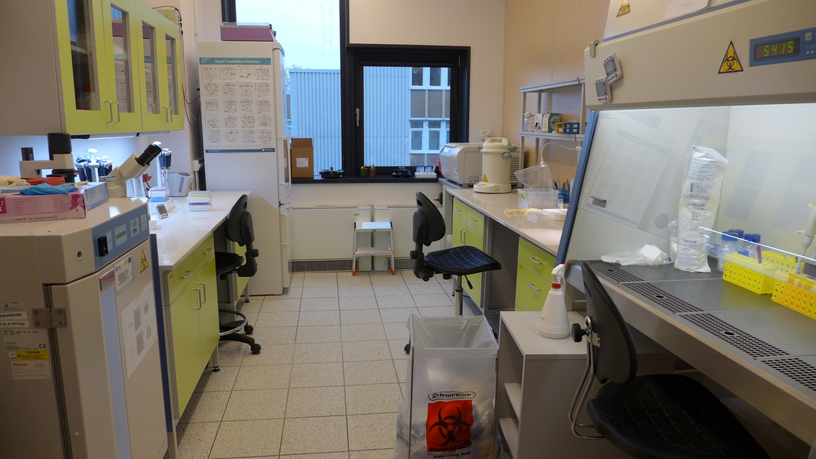
[[Image:Experimental_facility_cell_culture_room.jpg|link={{SERVER}}/images/b/bf/Experimental_facility_cell_culture_room.jpg||thumb|Tissue culture room.]] | [[Image:Experimental_facility_cell_culture_room.jpg|link={{SERVER}}/images/b/bf/Experimental_facility_cell_culture_room.jpg||thumb|Tissue culture room.]] | ||
*laminar hoods | *laminar hoods | ||
| Line 52: | Line 51: | ||
===Microscopy Unit=== | ===Microscopy Unit=== | ||
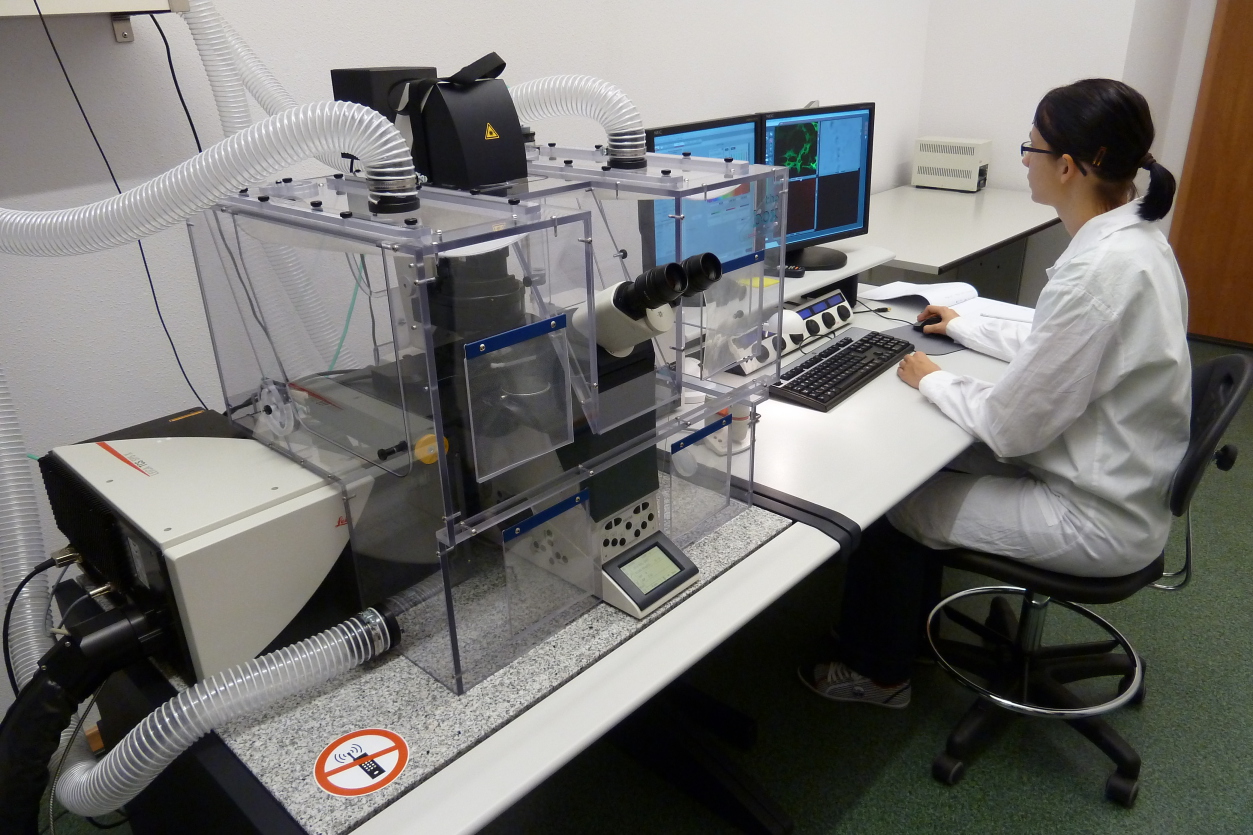
[[Image:Experimental_facility_microscopy_unit.jpg|link={{SERVER}}/images/3/37/Experimental_facility_microscopy_unit.jpg|thumb|Confocal microscopy unit.]] | [[Image:Experimental_facility_microscopy_unit.jpg|link={{SERVER}}/images/3/37/Experimental_facility_microscopy_unit.jpg|thumb|Confocal microscopy unit.]] | ||
* confocal microscope ''Leica TCS SP5 X'' with environmental chamber <div class="toccolours mw-collapsible mw-collapsed | * confocal microscope ''Leica TCS SP5 X'' with environmental chamber <div class="toccolours mw-collapsible mw-collapsed"><div class="mw-collapsible-content"> | ||
** based on inverted, fully automated microscope Leica DMI6000 | ** based on inverted, fully automated microscope Leica DMI6000 | ||
** environmental chamber with temperature, CO<sub>2</sub> and humidity control | ** environmental chamber with temperature, CO<sub>2</sub> and humidity control | ||
| Line 67: | Line 66: | ||
*** HeNe 633 nm | *** HeNe 633 nm | ||
** '''Objectives''': | ** '''Objectives''': | ||
*** HC PL FLUOTAR | *** HC PL FLUOTAR 5×/0.15 | ||
*** HC PL APO | *** HC PL APO 10×/0.40 CS | ||
*** HC PL APO | *** HC PL APO 20×/0.70 CS | ||
*** HCX PL APO | *** HCX PL APO 40×/0.85 CS | ||
*** HCX PL APO | *** HCX PL APO 63×/1.40-0.60 OIL CS | ||
** '''Fluorescence filter cubes''': | ** '''Fluorescence filter cubes''': | ||
*** GFP (filter "GFP"): excitation: BP470/40, dichroic: 495, emission: BP525/50 | *** GFP (filter "GFP"): excitation: BP470/40, dichroic: 495, emission: BP525/50 | ||
| Line 80: | Line 79: | ||
*** DAPI "band-pass" (filter "A4 ET"): excitation BP360/40, dichroic: 400, <br>emission: BP470/40</div></div> | *** DAPI "band-pass" (filter "A4 ET"): excitation BP360/40, dichroic: 400, <br>emission: BP470/40</div></div> | ||
* TIRF microscope ''Leica AM TIRF MC'' <div class="toccolours mw-collapsible mw-collapsed | * TIRF microscope ''Leica AM TIRF MC'' <div class="toccolours mw-collapsible mw-collapsed"><div class="mw-collapsible-content"> | ||
** based on inverted, fully automated microscope Leica DMI6000 with AFC | ** based on inverted, fully automated microscope Leica DMI6000 with AFC | ||
** environmental chamber with temperature, CO<sub>2</sub> and humidity control | ** environmental chamber with temperature, CO<sub>2</sub> and humidity control | ||
** methods: TIRF, FRET, fluorescence, bright filed, DIC, polarization | ** methods: TIRF, FRET, fluorescence, bright filed, DIC, polarization | ||
** fully integrated module for Multi-Color Total Internal Reflection Fluorescence | ** fully integrated module for Multi-Color Total Internal Reflection Fluorescence | ||
*** specimen penetration depth from 70 nm to 300 nm | *** specimen penetration depth from 70 nm to 300 nm | ||
*** lasers: 405, 488, 561 and 635 nm with AOTF | *** lasers: 405, 488, 561 and 635 nm with AOTF | ||
*** TIRF | *** TIRF objective: HCX PL APO 100×/1.47 OIL CORR TIRF | ||
** fluorescence light source: 120 W mercury metal halide lamp | ** fluorescence light source: 120 W mercury metal halide lamp | ||
** '''Cameras''': | ** '''Cameras''': | ||
*** EM-CCD, Hamamatsu C9100-02 ( | *** EM-CCD, Hamamatsu C9100-02 (1000×1000 px, monochrome, cooled, high EM gain, up to 256 fps, 31 fps at full resolution) | ||
*** Leica DFC365FX ( | *** Leica DFC365FX (1391×1040 px, monochrome, cooled, up to 122 fps, 21 fps at full resolution) | ||
*** Leica DFC295 ( | *** Leica DFC295 (2048×1536 px, color, up to 35 fps, 12 fps at full resolution) | ||
** '''Objectives''': | ** '''Objectives''': | ||
*** N PLAN 2. | *** N PLAN 2.5×/0.07 | ||
*** N PLAN | *** N PLAN 5×/0.12 | ||
*** HCX PL S-APO | *** HCX PL S-APO 10×/0.30 | ||
*** HCX PL FL L | *** HCX PL FL L 20×/0.40 CORR | ||
*** HCX PL FL L | *** HCX PL FL L 63×/0.70 CORR | ||
*** HCX PL APO | *** HCX PL APO 100×/1.47 OIL CORR TIRF</div></div> | ||
* confocal bio-imaging system: ''BD Pathway 435'' | * high-throughput microplate imager: ''Operetta CLS'' (PerkinElmer) | ||
* integrated system of | * confocal bio-imaging system: ''BD Pathway 435'' (BD Biosciences) | ||
* inverted fluorescent microscope ('' | * integrated system of atomic force microscopy (AFM), confocal microscopy and Raman spectroscopy (''NTEGRA Spectra'', NT-MDT Co.) | ||
* inverted fluorescent microscope (''DMI3000B'', Leica) | |||
* optical tweezers | * optical tweezers | ||
| Line 112: | Line 112: | ||
*microPIV platform: ''Nikon Eclipse E-50i'' epi-fluorescent microscope with double-shutter CCD camera and double-pulsed Nd:YAG laser | *microPIV platform: ''Nikon Eclipse E-50i'' epi-fluorescent microscope with double-shutter CCD camera and double-pulsed Nd:YAG laser | ||
*stereo microscopes | *stereo microscopes | ||
*high-precision syringe pumps (''neMESYS'', | *high-precision syringe pumps (''neMESYS'', Cetoni GmbH) | ||
*high-precision peristaltic pumps (''peRISYS'', | *high-precision peristaltic pumps (''peRISYS'', Cetoni GmbH) | ||
*syringe pumps, peristaltic pump, micro gear pump | *syringe pumps, peristaltic pump, micro gear pump | ||
*high-speed intensified CMOS camera (''Lambert HiCAM 500'') | *high-speed intensified CMOS camera (''Lambert HiCAM 500'') | ||
*high-speed camera (''Photron'', ''FASTCAM Mini AX100'' 540K-M-8GB) | *high-speed camera (''Photron'', ''FASTCAM Mini AX100'' 540K-M-8GB) | ||
*high-speed CMOS camera (''pco.1200 hs'', | *high-speed CMOS camera (''pco.1200 hs'', PCO IMAGING) | ||
*double-shutter CCD camera (''SensiCam'', | *double-shutter CCD camera (''SensiCam'', PCO IMAGING) | ||
*equipment for microfluidic chip fabrication by soft photolithography, ''i.a.'' spin coater, plasma cleaner, UV curing system, laboratory ovens, heating plates | *equipment for microfluidic chip fabrication by soft photolithography, ''i.a.'' spin coater, plasma cleaner, UV curing system, laboratory ovens, heating plates | ||
*equipment for microfluidic chip fabrication by micro-milling | *equipment for microfluidic chip fabrication by micro-milling | ||
| Line 136: | Line 136: | ||
===Other equipment=== | ===Other equipment=== | ||
*laser plotter equipped with Jasper X0-30 femtosecond laser (''Fluence'', Poland), designed for micromachining, cutting, and surface structuring of a wide range of materials (e.g., glass, plastics) | |||
*high-energy, double-pulsed Nd:YAG nanosecond laser (''EKSPLA NL303D'') | |||
*high frequency 1 kHz pulsed Nd:YAG laser (''EKSPLA NL202'') | |||
*double-pulsed Nd:YAG laser (''SoloPIV'', New Wave Research Inc.) | |||
*solenoid micro-valves | *solenoid micro-valves | ||
*pressure sensors | *pressure sensors | ||
*temperature sensors | *temperature sensors | ||
*accessories like camera lenses, microscope objectives, mirrors, lenses, halogen lamps, sensors of temperature, pressure, humidity, viscosity and others | *accessories like camera lenses, microscope objectives, mirrors, lenses, halogen lamps, sensors of temperature, pressure, humidity, viscosity and others | ||
<br /> | <br /> | ||
Latest revision as of 19:14, 11 December 2023
Our experimental facility consists of 11 rooms with the total area of ca. 200 m2. Each room is equipped with air conditioning, air humidity control system, and connected to an automatic stand-by power generator. Additionally, Tissue Culture Room, Molecular Biology Unit and Microfluidics Unit are equipped with HEPA H14 air filtration system.
We have the permission for the contained use of GMOs in our research.
Tissue Culture Unit
- laminar hoods
- CO2 incubators
- inverted laboratory microscope (Leica DM IL LED)
- automated cell counter (Bio-Rad TC10 Automated Cell Counter)
- water baths
- shaker
- incubated shaker
- centrifuge
- micro-centrifuge
- freezer −25 °C
- ultra-low temperature freezer −86 °C
- cryogenic storage Dewar
- autoclave 108L (HMC HG113)
- water purification system
- icemaker
- accessories: vortex, magnetic stirrer, heating block, aspirators
Molecular Biology Unit

- laminar hood
- CO2 incubator
- inverted laboratory microscope (Leica DM IL LED)
- bioanalyzer (Agilent 2100 Bioanalyzer)
- nucleoporator (Lonza Nucleofector IIS)
- PCR thermocycler (Eppendorf Mastercycler)
- real-time PCR (Applied Biosystems QuantStudio 12K Flex)
- digital PCR (Applied Biosystems QuantStudio 3D)
- flow cytometer (BD FACSCalibur)
- fluorometer (Thermo Scientific Fluoroskan Ascent Microplate Fluorometer)
- spectrophotometer (Thermo Scientific Multiskan GO Microplate Spectrophotometer)
- Western blot set
- nucleic acid gel separation system
- imaging system (Bio-Rad ChemiDoc MP Imaging System)
- gel imaging system (Bio-Rad Molecular Imager GelDoc XR+)
- centrifuge
- micro-centrifuge
- water baths
- refrigerator
- accessories: vortex, magnetic stirrer, balance, heating block
- dark-room (separate)
Microscopy Unit
- confocal microscope Leica TCS SP5 X with environmental chamber
- based on inverted, fully automated microscope Leica DMI6000
- environmental chamber with temperature, CO2 and humidity control
- scanning stage with Super-Z Galvo
- 3 PMT confocal detectors
- 1 brightfield detector
- digital camera Leica DFC365FX (cooled, 21 fps at full resolution 1392×1040)
- fluorescence light source: 120 W mercury metal halide lamp
- Lasers:
- White Light Laser (WLL): tunable excitation wavelengths from 470 nm to 670 nm
with 1 nm resolution, up to 8 excitation lines can be used simultaneously - diode 405 nm
- multi-wavelengths Argon laser: 458, 476, 488, 496 and 514 nm
- DPSS 561 nm
- HeNe 633 nm
- White Light Laser (WLL): tunable excitation wavelengths from 470 nm to 670 nm
- Objectives:
- HC PL FLUOTAR 5×/0.15
- HC PL APO 10×/0.40 CS
- HC PL APO 20×/0.70 CS
- HCX PL APO 40×/0.85 CS
- HCX PL APO 63×/1.40-0.60 OIL CS
- Fluorescence filter cubes:
- GFP (filter "GFP"): excitation: BP470/40, dichroic: 495, emission: BP525/50
- FITC (filter "I.3"): excitation BP450-490, dichroic: 510, emission: LP515
- TRITC (filter "N2.1"): excitation BP515-560, dichroic: 580, emission: LP590
- Red "band-pass" (filter "N3 ET"): excitation BP546/11, dichroic: 565,
emission:BP 600/40 - DAPI "long-pass" (filter "A"): excitation BP340-380, dichroic: 400, emission: LP425
- DAPI "band-pass" (filter "A4 ET"): excitation BP360/40, dichroic: 400,
emission: BP470/40
- TIRF microscope Leica AM TIRF MC
- based on inverted, fully automated microscope Leica DMI6000 with AFC
- environmental chamber with temperature, CO2 and humidity control
- methods: TIRF, FRET, fluorescence, bright filed, DIC, polarization
- fully integrated module for Multi-Color Total Internal Reflection Fluorescence
- specimen penetration depth from 70 nm to 300 nm
- lasers: 405, 488, 561 and 635 nm with AOTF
- TIRF objective: HCX PL APO 100×/1.47 OIL CORR TIRF
- fluorescence light source: 120 W mercury metal halide lamp
- Cameras:
- EM-CCD, Hamamatsu C9100-02 (1000×1000 px, monochrome, cooled, high EM gain, up to 256 fps, 31 fps at full resolution)
- Leica DFC365FX (1391×1040 px, monochrome, cooled, up to 122 fps, 21 fps at full resolution)
- Leica DFC295 (2048×1536 px, color, up to 35 fps, 12 fps at full resolution)
- Objectives:
- N PLAN 2.5×/0.07
- N PLAN 5×/0.12
- HCX PL S-APO 10×/0.30
- HCX PL FL L 20×/0.40 CORR
- HCX PL FL L 63×/0.70 CORR
- HCX PL APO 100×/1.47 OIL CORR TIRF
- high-throughput microplate imager: Operetta CLS (PerkinElmer)
- confocal bio-imaging system: BD Pathway 435 (BD Biosciences)
- integrated system of atomic force microscopy (AFM), confocal microscopy and Raman spectroscopy (NTEGRA Spectra, NT-MDT Co.)
- inverted fluorescent microscope (DMI3000B, Leica)
- optical tweezers
Microfluidics Unit

- microfluidic platform for cell culturing CellASIC ONIX
- microPIV platform: Nikon Eclipse E-50i epi-fluorescent microscope with double-shutter CCD camera and double-pulsed Nd:YAG laser
- stereo microscopes
- high-precision syringe pumps (neMESYS, Cetoni GmbH)
- high-precision peristaltic pumps (peRISYS, Cetoni GmbH)
- syringe pumps, peristaltic pump, micro gear pump
- high-speed intensified CMOS camera (Lambert HiCAM 500)
- high-speed camera (Photron, FASTCAM Mini AX100 540K-M-8GB)
- high-speed CMOS camera (pco.1200 hs, PCO IMAGING)
- double-shutter CCD camera (SensiCam, PCO IMAGING)
- equipment for microfluidic chip fabrication by soft photolithography, i.a. spin coater, plasma cleaner, UV curing system, laboratory ovens, heating plates
- equipment for microfluidic chip fabrication by micro-milling
- 3D printer (Zortrax Inventure)
Biomaterials Unit
- setup for polymer nanofibers fabrication by electrospinning equipped with environmental chamber
- 3D bioprinter (Cellink, INKREDIBLE S-10002-001)
- lyophilizator (LABCONCO, FreeZone 2.5 Liter -84C Benchtop Freeze Dryer 710201030)
- plasma cleaner (Diener, ZEPTO PCCE)
- fume hood
- accessories: analytical balances, centrifuges, vortexes, magnetic stirrers, blood roller mixer, water baths, refrigerated/heating circulators, conductometer, pH-meter, vacuum pump, pipettes, etc.
Other equipment
- laser plotter equipped with Jasper X0-30 femtosecond laser (Fluence, Poland), designed for micromachining, cutting, and surface structuring of a wide range of materials (e.g., glass, plastics)
- high-energy, double-pulsed Nd:YAG nanosecond laser (EKSPLA NL303D)
- high frequency 1 kHz pulsed Nd:YAG laser (EKSPLA NL202)
- double-pulsed Nd:YAG laser (SoloPIV, New Wave Research Inc.)
- solenoid micro-valves
- pressure sensors
- temperature sensors
- accessories like camera lenses, microscope objectives, mirrors, lenses, halogen lamps, sensors of temperature, pressure, humidity, viscosity and others
Laboratory equipment was partially financed by the European Union – the European Regional Development Fund within the Operational Programme “Innovative Economy” for 2007–2013.